The STANDARD ultrasound examination of the fetus has been agreed upon between three professional organizations, the American Institute of Ultrasound in Medicine (AIUM)) (2003), the American College of Radiology (ACR) (2003), and the American College of Obstetricians and Gynecologists (ACOG) (2004). All three organizations recommend identical protocols for imaging the fetus; irrespective of whether the examination is performed by a radiologist, obstetrician, or specialist in fetal-maternal medicine. The section on Screening Ultrasound covers all of the components of the STANDARD ultrasound examination in more detail. This section will focus on the examination of the fetal heart.
When ultrasound was first used in the early 1980's to identify the fetal heart investigators were uncertain as to the approach that should be used to identify congenital heart defects. During this time period Dr. DeVore was one of the early pioneers in the development of this technology (see Dr. DeVore's publications). One of the difficulties, however, was how to approach examination of the fetal heart so that those not experienced in Fetal Echocardiography could identify heart defects. In 1985 Dr. DeVore published one of the first papers suggesting that abnormalities of the four-chamber view of the heart could identify fetuses with heart defects. This was followed by numerous studies confirming this observation made by Dr. DeVore. The STANDARD examination of the heart is as follows:
The basic cardiac examination includes a 4-chamber view of the fetal heart. If technically feasible, an extended basic cardiac examination can also be attempted to evaluate both outflow tracts.
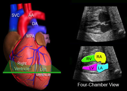
What is the Four-Chamber View of the Heart?
When the ultrasound beam is directed perpendicular to the chest of the fetus four-chambers of the fetal heart are identified. These chambers consist of the right and left atrial and ventricular chambers, with their respective valves that connect the atrial with the ventricular chambers. In 1985 Dr. DeVore suggested that, because of fetal circulation, abnormal cardiac anatomy could alter the way in which the four-chamber view appeared. A number of subsequent studies validated this concept. The following images illustrate the ultrasound approach used to examine the four-chamber view.
Imaging the four-chamber view is accomplished by directing the ultrasound beam perpendicular to the fetal chest. At this level, the four-chamber view is identified. This view contains the right atrium (RA), left atrium (LA), right ventricle (RV) and left ventricle (LV).
This is the four-chamber view of the fetal heart.
Examples of Abnormalities of the Four-Chamber View
The following are examples of abnormalities of the four-chamber view. To view the beating heart, click on the play button for each video clip.
Normal 4-Chamber View
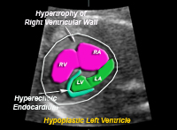
Hypoplastic left ventricle. This is the result of underdevelopment of the left ventricular chamber, left atrial chamber, mitral valve and aortic valve.
Labeled Image of the hypoplastic left ventricle. RV=right ventricle, LV=left ventricle, RA=right atrium, LA=left atrium
Normal 4-Chamber View
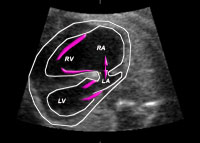
This is an example of an abnormal tricuspid valve of the right ventricle. The valve is displaced low in the right ventricular chamber. This results in changes in the four-chamber view that include a larger right atrium, smaller left atrium, and abnormal valve motion of the tricuspid valve.
Labeled Image of the hypoplastic left ventricle. RV=right ventricle, LV=left ventricle, RA=right atrium, LA=left atrium
Normal 4-Chamber View
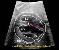
This is an example of an endocardial cushion defect, often seen in fetuses with Down syndrome. When this is present there is a ventricular and atrial septal defect which means that a portion of the wall dividing the left and right sides of the heart is missing.
This is an endocardial cushion defect. The arrows demonstrate the absence of the ventricular and atrial septum. There is also fluid around the edge of the heart (PE) that should not be present. LV=left ventricle, LA=left atrium, RV=right ventricle, RA=right atrium
Limitations of the Four-Chamber View
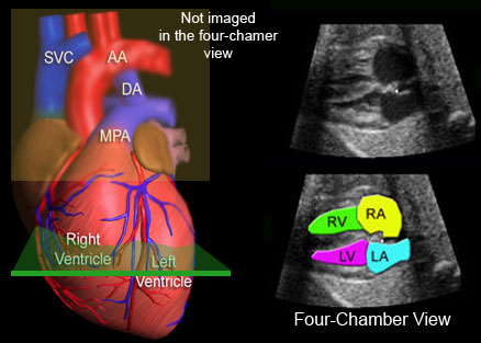
Although the four-chamber view is useful for identifying abnormalities of the fetal heart, several defects may always demonstrate an abnormal four-chamber view. The reason for this is because heart defects may only involve the outflow tracts. The outflow tracts consist of the main pulmonary artery exiting the right ventricle and the aorta exiting the left ventricle. The reason that the four-chamber view does not identify these malformations is because the image of the four-chamber view is obtained at the level of the ventricles and atria, and not at the levels of the outflow tracts.
This illustrates the level that the four-chamber view is imaged (green). The main pulmonary artery (MPA), ductus arteriosus (DA), and the aorta (AA) are not imaged at the level of the four-chamber view. SVC=superior vena cava, RV=right ventricle, RA=right atrium, LV=left ventricle, LA=left atrium.
Examination of the Outflow Tracts
There are several approaches that the examiner can use to identify the outflow tracts of the heart that use the four-chamber view as the initial reference image.
Rotational Technique
This technique was first described by Dr.DeVore in 1992. After imaging the four-chamber view, the physician rotates and then rocks the transducer to image the ascending aorta and the main pulmonary artery. This maneuver identifies that (1) the main pulmonary artery and aorta are perpendicular to each other as they exit their respective ventricles, (2) these vessels are similar in size, and (3) the aortic and pulmonary valves are normal, and (4) the aortic arch.
Movement of the transducer used to acquire the views using the Rotational Technique.
The Rotation Technique.
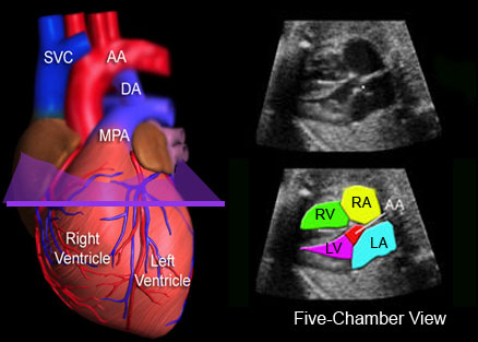
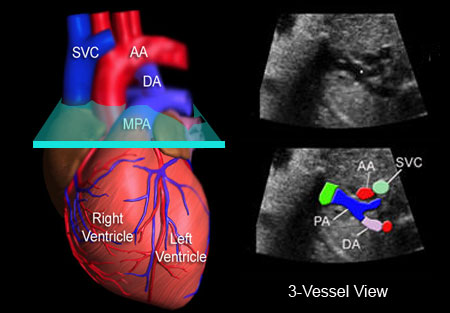
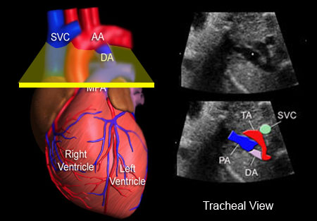
Sweep Technique
This technique was described by Yoo et al and Yagel et al and involves sweeping the transducer beam in a transverse plane from the level of the four-chamber view towards the fetal neck. By doing so, the outflow tract vessels are observed. The sweep consists of the following views: four-chamber view, five-chamber view, main pulmonary artery or 3-vessel view, and the tracheal view.
.
This illustrates movement of the transducer for obtaining the Sweep image sequences described above.
This video clip illustrates the Sweep Technique from the stomach to the fetal neck.
The following images illustrate the four different levels used to identify cardiac anatomy using the Sweep Technique.
Four-Chamber View
This illustrates the level that the four-chamber view is imaged (green). The main pulmonary artery (MPA) and the aorta (AA) are not imaged at this level. SVC=superior vena cava, DA=ductus arteriosus, RV=right ventricle, RA=right atrium, LV=left ventricle, LA=left atrium.
Five-Chamber View
This is the level of the five-chamber view and illustrates the aorta (AA) exiting the left ventricle. RV=right ventricle, RA=right atrium, LV=left ventricle, LA=left atrium.
3-vessel View
This is the 3-vessel view because 3 vessels are observed; the superior vena cava (SVC), the cross-section of the ascending aorta (AA), and the full length of the main pulmonary artery (PA). This is an important view because it illustrates the ascending aorta perpendicular to the main pulmonary artery. If these two vessels are not perpendicular to each other at this level, then a serious heart defect, transposition of the great vessels, is most likely present. DA=ductus arteriosus.
Tracheal View
This is the tracheal view because these vessels are at the level of the trachea. This view is important because it illustrates the transverse aortic arch (TA) and the ductus arteriosus (DA) merging with the thoracic aorta.
Summary of All Views
The four-chamber view and the outflow tracts of the heart are imaged by first locating the four-chamber view of the heart and then sweeping the transducer towards the fetal neck. When this is done, the five-bomber, 3-vessel, and tracheal views are identified.
Examples of Pathology
The following are examples of heart abnormalities that are identified using the sweep technique.
This is an example of Transposition of the Great Arteries. When this is present the left ventricle gives rise to the pulmonary artery and the right ventricle to the aorta. Because of fetal circulation this creates no problem for the fetus. However, once birth occurs, the blood flow to the lungs and body is abnormal. Newborns with this condition require surgery to survive.
This illustrates the pathology. The aorta (A) exits the right ventricle (RV) and the main pulmonary artery (MPA) exits the left ventricle. This image is taken at the level of the 5-chamber view. Both the aorta and main pulmonary artery are parallel to each other instead of being perpendicular.
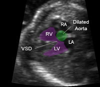
This is an example of Tetralogy of Fallot. There is a ventricular septal defect that straddles the dilated aorta. This defect is not usually seen in the four-chamber view.
This illustrates the pathology. The aorta is increased in size and straddles the ventricular septal defect.