Use of 3D Ultrasound

Fetuses with Down syndrome have multiple malformations, as has been previously discussed. Of the many defects, 3D/4D ultrasound has been useful in evaluating the skeletal and cardiovascular systems in fetuses with this condition. This section will describe some examples of 3D ultrasound used to examine fetal anatomy in fetuses at risk for Down syndrome. The 3D/4D ultrasound often enhances the interpretation of fetal anatomy when the 2D image is difficult to interpret. One of the limitations of 3D/4D ultrasound is that imaging is dependent upon several factors that include the gestational age, position of the fetus within the uterus, whether the mother has excessive adipose or fatty tissue, and the amount of amniotic fluid surrounding the fetus. The following images are examples of fetal anatomy under ideal imaging conditions. The images displayed on this page may not be obtainable in all examinations.
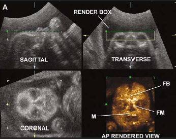
FACE
A number of studies have reported the association between an absent or small nasal bone and an increased risk for Down syndrome. The short or absent nasal bone has also been associated with Trisomy 18, a lethal chromosomal defect, as well as other malformations. In the section on Real-Time B-Mode ultrasound facial profiles of the nasal bone showed this pathology. If the 2D ultrasound does not demonstrate two nasal bones, then 3D ultasound may be useful. For example, a fetus with Down syndrome can have one nasal bone that appears normal, and the second bone hypoplastic or absent. For this reason, 3D ultrasound reconstruction of the nasal bone and other facial bones is useful. Click this LINK to view abstracts from the medical literature.
|

|

|

|
SPINE
Several studies (Edwards et al, Willich et al., and Kriss) have found that fetuses with Down syndrome are either missing or have an underdeveloped 12th rib. Using 2D ultrasound it is not possible to examine the ribs of the fetus and determine their length. This is because of their curved nature. However, with 3D ultrasound the ribs can be examined and their number determined.
|

HEART
Three and 4D ultrasound allows the physician to simultaneously examine multiple views of the heart and vessels as well as evaluate the heart in a 3D and 4D model. These examples illustrate pathology observed in fetuses with Down syndrome.