Genetic Ultrasound in Women 35 and Older

Can Ultrasound Be Used To Identify Fetuses With Down Syndrome?
Early Ultrasound Studies
Until recently, medical studies in which ultrasound evaluation of fetal anatomy has been used to identify fetuses with Down syndrome have focused on individual features of Down syndrome. Individual ultrasound markers which have been studied include the length of the middle bone of 5th digit of the hand, the length of the femur (upper leg bone), the length of the humerus (upper arm bone), length of the foot, the presence of a thick pad of skin behind the neck (nuchal skin fold), the appearance of the kidneys, and bowel. While these studies have been helpful, they did not demonstrate a detection rate for Down syndrome which was greater than maternal Triple Marker serum screening. In addition, no studies focused on examination of the fetal heart, the most frequent birth defect in infants with Down syndrome. The reason for this was because the fetal heart was the most difficult part of the fetal ultrasound examination to perform and interpret.
Color Doppler Ultrasound To Detect Down Syndrome
In 1990, a study was initiated Dr. DeVore, in collaboration with Dr. Omar Alfi, a Pediatric Geneticist who was the former director of Alfigen, The Genetics Institute, (Pasadena, California),where the study was conducted. The purpose of the study was to determine whether color Doppler ultrasound evaluation of the cardiovascular system could be used to improve the detection rate for Down syndrome as well as other chromosomal abnormalities. The results of the original study were published in the medical journal Obstetrics and Gynecology, the official journal of The American College of Obstetricians and Gynecologists. The results of the study found that examination of the fetus with real-time ultrasound identified 30% of fetuses with Down syndrome, a number which was consistent with prior studies in which multiple organ systems were examined. However, the use of color Doppler ultrasound increased the yield from 29% to 87%. In addition, the detection rate of all abnormal chromosomes, of which Down syndrome is the most common, increased from 36% to 75%. If the patients who were obese in which ultrasound examination was inadequate were removed from the analysis, the detection rate for Down syndrome increased from 87% to 93%.
Following the initial report by Dr. DeVore, a more comprehensive study was published in 2000 in the journal of Ultrasound in Obstetrics and Gynecology in which 80 fetuses with Down syndrome and 2,000 controls were examined. This study demonstrated the following:
1. Using a B-Mode ultrasound, without examining the fetal heart, detected 60 of fetuses with Down syndrome.
2. Using B-Mode ultrasound and evaluating the four-chamber view of the heart for disproportion, 75% of fetuses with Down syndrome were detected.
3. Using B-Mode ultrasound plus color Doppler ultrasound to examine the fetal heart in greater detail identified 91% of fetuses with Down syndrome.
4. Genetic Ultrasound, when practiced by physicians with special training in this area, is a useful tool to adjust the risk for Down syndrome.
At the present time Genetic Ultrasound is an accepted methodology that can be used to determine the risk for Down syndrome. A recent publication from the American College of Obstetricians and Gynecologists entitled the ACOG Practice Bulletin, “Ultrasound in Pregnancy” (Num 58, December 2004), states:
“A second-trimester specialized ultrasound examination may be targeted to detect fetal aneuploidy (Down syndrome). This type of examination has been offered in some centers for the past several years and is aimed at the detection of a range of minor anatomic features associated with an increased risk for fetal aneuploidy…. The standard examination is less likely to detect the minor anatomic features associated with aneuploidy.”
The Use of Genetic Ultrasound to Decrease the Risk for Down Syndrome in Women 35 Years of Age and Older
Genetic Ultrasound can be used in two clinical situations in women 35 years of age and older. With the inception of maternal serum screening using the Triple Marker test and more recently the QUAD test, approximately 25% of women 35 years of age and older will be identified to be at high risk for Down syndrome. Many of these women do not desire genetic amniocentesis and often ask if another non-invasive test could be performed to re-evaluate their risk. Genetic Ultrasound fit this role perfectly.
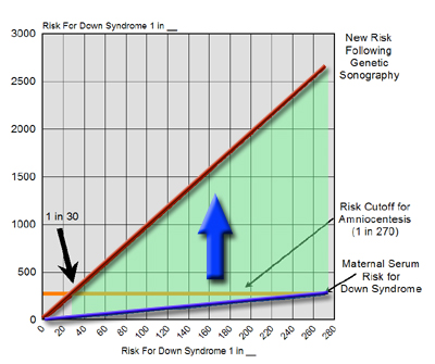
The reduction in risk depends upon how accurate the Genetic Ultrasound detects Down syndrome. For example, if B-Mode and color Doppler are used, the detection rate of Down syndrome is 91%, as described above. Therefore, if a woman 35 years of age or older is informed that she has an increased risk for Down syndrome following maternal serum screening, then her final risk can be reduced if the Genetic Ultrasound is normal. The following graph illustrates the reduction in risk for Down syndrome based an abnormal risk following maternal serum screening.
|
The Use of Genetic Ultrasound to Increase the Detection Rate for Down Syndrome in Women 35 Years of Age and Older
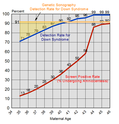
Genetic Ultrasound also has several advantages over Triple Marker screening. Although the medical literature states that the "overall" detection rate for Down syndrome in women between 35 and 45 is 89%, this rate varies as a function of maternal age. The following graph illustrates this point. Women at age 35 only have a 71% detection rate for Down syndrome, not 89%. The reason for this is because only 13% are screen-positive. Not until age 40 does the detection rate for Down syndrome approach 89%. However, the detection rate for Down syndrome using Genetic Sonography is 91%, irrespective of maternal age. Therefore, Genetic Ultrasound is more advantageous for detecting Down syndrome than maternal serum Triple Marker screening for women between ages 35 and 40.
|
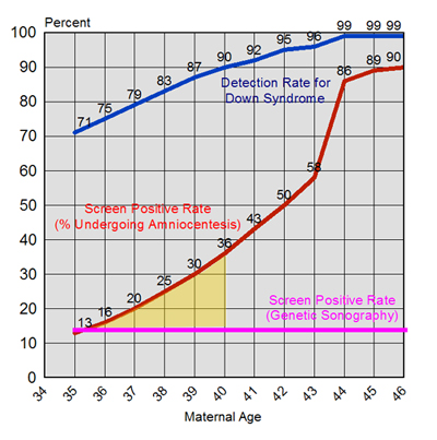
For women between 35 and 40 years of age the risk of pregnancy complications is higher for Triple Marker testing than it is for Genetic Ultrasound, with a lower yield for detecting this condition. Therefore, not only is the detection rate for Down syndrome higher using Genetic Ultrasound, the amniocentesis rate is lower (14%) than for Triple Marker testing (13% to 36%).
|
The concept of using Genetic Ultrasound in women who tested negative following Triple-Marker screening was analyzed by Dr.DeVore and Dr. Roberto Romero of the Perinatal Research Branch of the National Institutes of Health and Human Development. They found that if all women who tested negative underwent Genetic Ultrasound, the detection rate for Down syndrome would increase to 98%, and was a safe alternative to universal amniocentesis.
|
